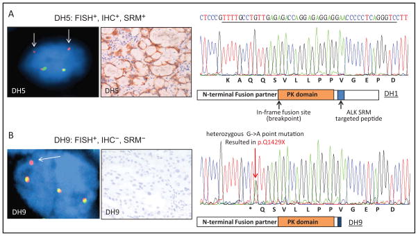
Fig. 3. Comparison of SRM with ALK FISH, IHC, and DNA sequencing in selective cases.
FISH and paired IHC for patient DH5 to represent FISH+/IHC+ cases; DNA sequence within the MS targeted peptide from DH1 to represent wild-type sequence. (A), ALK FISH testing shows deletion of the 5′ (green) signal with retained 3′ (orange) signal, consistent with rearrangement. Arrows indicate the rearranged red signal. (B), The FISH+, SRM−, and IHC− case (DH9) showed a nonsense point mutation that resulted in a stop codon (p.Q1429X); therefore, nonfunctional fusion protein would be produced.

