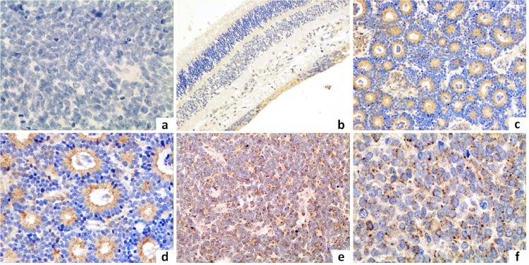
Fig 1. Immunohistochemistry in retinoblastoma tissues.
1a: Negative control stained with non-specific immunoglobulin; 1b: Immunostaining of AEG-1 in the relatively normal retinal tissue of the peri-tumor region (original magnification, ×100). 1c, d: Immunostaining of AEG-1 in well-differentiated specimens (original magnification, c: ×200; d: ×400); 1e, f: Immunostaining of AEG-1 in poorly-differentiated specimens (original magnification e: ×200; f: ×400).

