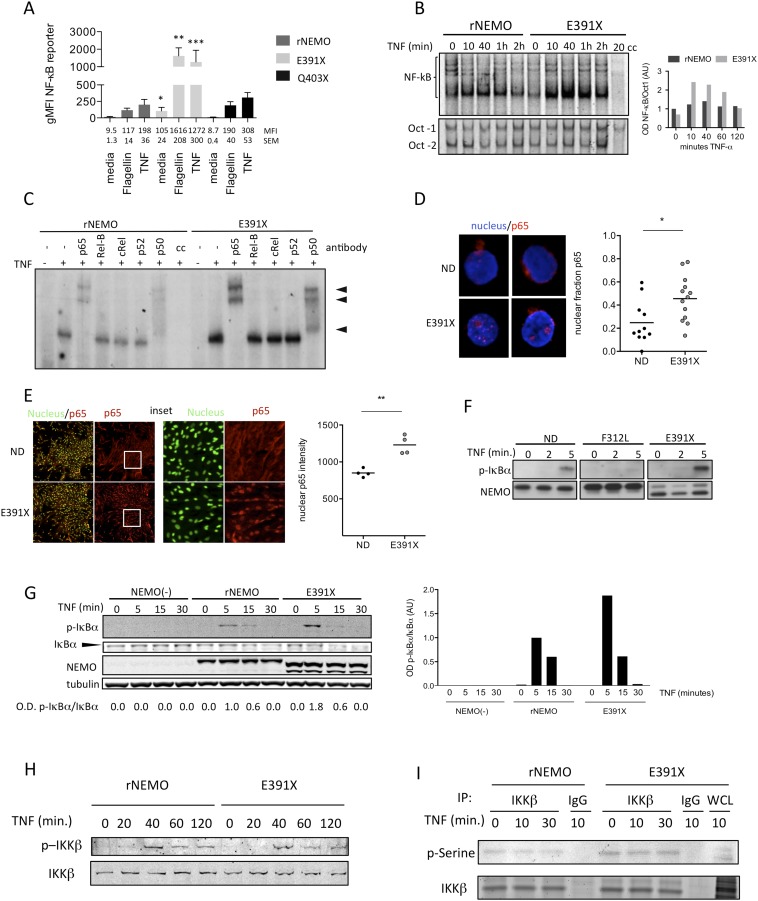
Fig. S3.
The canonical NF-κB pathway is activated in NEMO and ΔCT-mutant reconstituted cells following TNF stimulation. (A) Quantitation of surface protein expression (n = 3) of Thy1.1 NF-κB reporter as done in Fig. 2B. Average geometric mean fluorescence intensity (gMFI) and SEM appear beneath each condition. (B) Nuclear extracts were incubated with labeled NF-κB consensus sequence oligonucleotide or control (Oct-1) sequence and visualized by EMSA. Nuclear extracts loaded in lane labeled “cc” were preincubated with 100-fold excess of unlabeled consensus NF-κB or Oct-1 oligonucleotide before incubation with labeled oligonucleotide. Densitometry of the NF-κB/DNA complexes normalized to Oct-1 from a representative experiment (n = 3). (C) Reconstituted Jurkat T cells were stimulated with TNF and specificity of nuclear NF-κB complexes was determined by antibody-induced supershift of DNA-protein complexes. (D) Healthy control and E391X-NEMO patient PBMC were isolated. Nuclei were stained followed by intracellular p65 staining on cells following fixation. Images of two representative unstimulated cells are shown. Quantitation indicates nuclear fraction of p65 determined from images obtained by confocal microscopy at 63× magnification of ∼10 cells per condition. (E) Healthy control and E391X-NEMO patient iPS-derived fibroblast-like mesenchymal stem cells in culture. Nuclear and intracellular p65 staining was performed on fixed cells. Images at 4× magnification of a representative area of unstimulated cells are shown. The area bounded by the white box is shown enlarged at 10× magnification in panels on the right. For quantitation, each circle represents a single experimental replicate and each replicate consists of images containing ∼2,000 cells (n = 2). (F) Activated NEMO-E391X and NEMO-F312L patient T cells were stimulated with TNF. Western blot of whole cell lysates was probed with antibody specific to phosphorylated serine residues 32 and 36 of IκBα, NEMO was probed as a loading control. The result shown is representative of three independent experiments. (G) Reconstituted Jurkat T cells were stimulated with TNF for the indicated times and detection of p-IkBa and IkBa was performed by Western blot (n = 2); optical densitometry indicating the ratio of p-IkBa/IkBa was done (Right). (H) Reconstituted Jurkat T cells were stimulated with TNF and detection of IKK-b and IKK-a serine phosphorylation was performed by specific antibody following Western blot. (I) Reconstituted Jurkat T cells were stimulated with TNF followed by immunoprecipitation of IKK-β and subsequent detection of total phospho-serine using a specific antibody.

