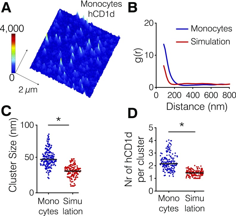
Fig. S3.
CD1d forms nanoclusters on the cell membrane of human monocytes. (A) Representative STED image of hCD1d on the cell membrane of monocytes. (B) Comparison between the correlation functions of the experimental data (blue) and corresponding Monte Carlo simulations (red) of hCD1d molecules randomly organized on the cell membrane using the same particle density as the experimental data. (C) Comparison between the experimental hCD1d cluster size (blue) and simulations of random organization (red). (D) Comparison between the number (Nr) of molecules per cluster obtained on the experimental data (blue) and the simulations (red). The larger values in cluster size and the number of molecules per cluster experimentally obtained compared with simulated data of random organization confirm that hCD1d is organized in nanoclusters on the surface of blood-derived primary hCD14+ monocytes. Experimental and simulated data are representative from at least 52 different STED images of 3 × 3 µm in size belonging to at least two different experiments. *P < 0.0001 (Student’s t test).

