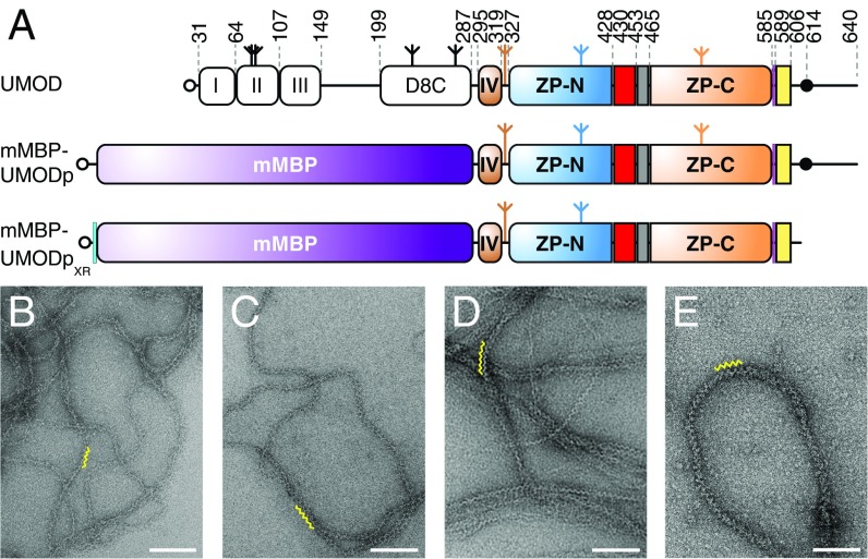
Fig. 1.
mMBP-fused UMODp forms filaments like native urinary UMOD. (A) Domain organization of urinary UMOD and recombinant constructs mMBP-UMODp and mMBP-UMODpXR. EGF domains are indicated by roman numerals. EGF IV identified by this study (brown), ZP-N/ZP-C linker (red), IHP (gray), CCS (magenta), CTP (yellow), and 6His-tag (cyan) are shown. Open circles, inverted tripods, and closed circles represent signal peptides, N-glycans, and GPI anchors, respectively. Electron micrographs of filaments of purified urinary UMOD (B), recombinant full-length UMOD from Madin–Darby canine kidney (MDCK) cells (C), purified elastase-digested urinary UMOD (D), and recombinant mMBP-UMODp from HEK293T cells (E). Yellow squiggles in B–E indicate the zigzag arrangement of UMOD repeats, which is most evident in samples lacking the N-terminal EGF I–III/D8C region. (Scale bars, 100 nm.)

