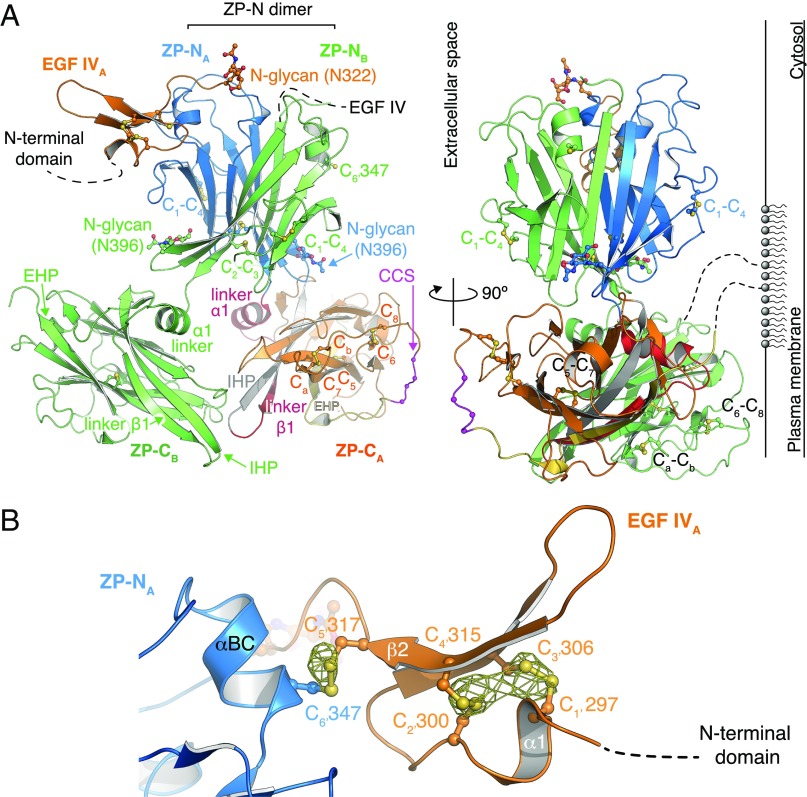
Fig. 2.
Structure of the protease-resistant core of human UMOD. (A) Overall UMODpXR architecture, with molecule A colored as in Fig. 1A and molecule B in green. N-glycans and Cys are depicted in a ball-and-stick representation. (Right) Possible orientation relative to the plasma membrane due to GPI anchoring is depicted. (B) Close-up view of EGF IV and its connection to ZP-N. An anomalous difference map calculated with Bijvoet differences collected at λ = 1.8 Å and contoured at 3.5 σ is shown as a yellow mesh.

