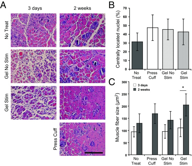
Fig. 3.
Ferrogel stimulation leads to improved muscle regeneration. (A) Histological cross-sections of tibialis anterior muscles stained with H&E 3 d and 2 wk after no treatment (No Treat), treatment with a pressure cuff (Press Cuff), treatment with a nonstimulated biphasic ferrogel (Gel No Stim), or treatment with a stimulated biphasic ferrogel (Gel Stim). (Scale bar, 100 μm.) (B) Quantification of myofibers residing in the defect containing centrally located nuclei 2 wk posttreatment. Values are expressed as a percentage of the total number of myofibers in the defect. (C) Quantified mean muscle fiber size in the defect area 3 d and 2 wk posttreatment. Data were compared using ANOVA with Bonferroni's post hoc test (n = 5; *P < 0.05). Error bars represent SDs.

