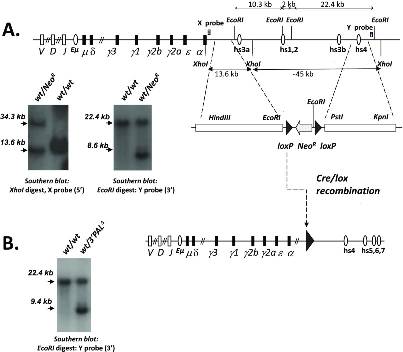
Fig. S1.
Targeting the IgH 3′RR proximal (quasi-palindromic) module in the mouse. (A) ES cell targeting strategy used to generate the 3′PAL KO mouse model in which the hs3a to hs3b region (so called 3′RR proximal module) was disrupted by insertion of a loxP site. Map of the mouse wt IgH 3′RR. Closed circles stand for transcriptional enhancers. Targeting construct and Southern blots of knockout ES cells or animals carrying NeoR insertion. The 5′ probe (X, 0.8-kb EcoRI-HindIII fragment) detects genomic 13.6-kb and 34.3-kb XhoI bands after homozygous recombination. The 3′ probe (Y, 0.6-kb XhoI-HindIII fragment) detects genomic 22.4-kb and 8.6-kb EcoRI bands in the targeted locus. (B) Map of the cre-deleted targeted locus. Southern blot on mouse tail DNA with 3′ Y probe detects genomic 22.4-kb and 9.4-kb EcoRI bands in the targeted locus.

