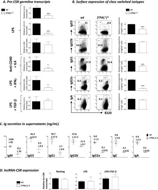
Fig. S4.
Impaired CSR in in vitro-stimulated B cells lacking the IgH 3′RR proximal module. Resting splenic B cells (CD43− fraction) from wt and 3′PALΔ/Δ mice were stimulated in vitro to induce CSR with LPS (IgG2b and IgG3), LPS + TGF-β (for IgA), LPS + IFN-γ (for IgG2a) or anti-CD40 + IL-4 (for IgG1 and IgE). (A) For each constant gene, germ-line transcription (Ix-Cx) was quantified by qRT-PCR and normalized to Gapdh expression after 48–72 h stimulation. Mean and SEM are reported, significant differences were indicated by P values: *P < 0.05, ***P < 0.001 according to the Mann–Whitney u test (n = 4–11 animals per genotype). (B) At day 4, percentage of isotype switched cells was analyzed by flow cytometry for surface Ig isotype expression: Center shows one representative experiment, Right displays statistical analysis of the proportion in vitro-induced isotype switched B cells for each condition. Mean and SEM are reported, significant differences are indicated by P values: *P < 0.05, ***P < 0.001 according to the Mann–Whitney u test (n = 12–28 animals per genotype). (C) Antibody isotype secretion was quantified by ELISA in stimulated B-cell supernatants at day 4. (D) Transcription of lncRNA for CSR was quantified by qRT-PCR (38) and normalized to Gapdh expression in wt and 3′PALΔ/Δ resting splenic and activated B cells for 48 (LPS) to 72 h (LPS+TGF-β). Mean and SEM are indicated (n = 6 animals).

