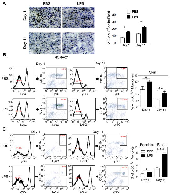
Figure 2. Super-low dose LPS increases recruitment of pro-inflammatory monocytes into wound tissues.
(A) Immunohistochemical staining of MOMA-2+ cells (brown color) around the skin wound at day 1 and day 11 after puncture, with quantification (n = 7). (B) Flow cytometry of Ly6G−/CD11b+/Ly6Chigh inflammatory monocytes in wound tissues after puncture. The frequency of inflammatory monocytes among total leukocytes was quantified. Data are shown from PBS- and super-low dose LPS-conditioned mice (day 1, n=6; day 11, n=7). (C) Flow cytometry of circulating Ly6G−/CD11b+/Ly6Chigh inflammatory monocytes. Data are shown from PBS- and super-low dose LPS-conditioned mice (day 1, n=6; day 11, n=7). Error bars show means ± s.e.m.; * P < 0.05; ** P < 0.01; *** P < 0.001; student t-test.

