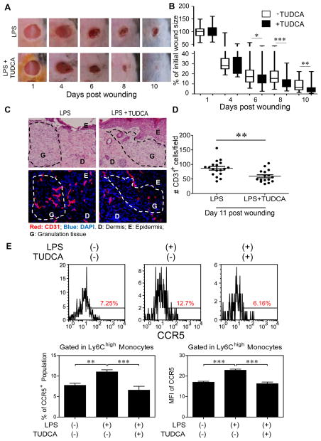
Figure 6. TUDCA facilitates wound healing in vivo.
Two groups of mice were injected i.p. with super-low dose LPS (5 ng/kg body weight) three times for 10 days before skin puncture and once every three days after puncture. After puncture, one group of mice was injected i.p. with TUDCA (5 mg/kg) daily for 10 days. (A) Wounds were monitored daily. Wound areas (% initial wound size, n>8) over the total observation periods in a box and whisker plot (B). Student t test, *, P < 0.05; **, P < 0.01; ***, P < 0.001. (C) H&E (C, top panel) and immuno-histochemical (C, bottom panel) staining of CD31-positive endothelial cells in skin as measurements of blood vessel sprouting within the wound (n=6 fields per slide, 6–7 slides per group). (D) Dot plot representing number of CD31-positive cells per viewing field. Error bars represent means ± s.e.m. Student t test, **, P < 0.01. (E) Peripheral blood cells were collected and CCR5 within Ly6G−/CD11b+/Ly6Chigh inflammatory monocytes was analyzed by flow cytometry. The frequency of CCR5+ population and MFI of CCR5 within Ly6Chigh inflammatory monocytes were quantified (n>5). Error bars show means ± s.e.m.; ns, not significant; * P < 0.05; **, P < 0.01; ***, P < 0.001; one-way ANOVA.

