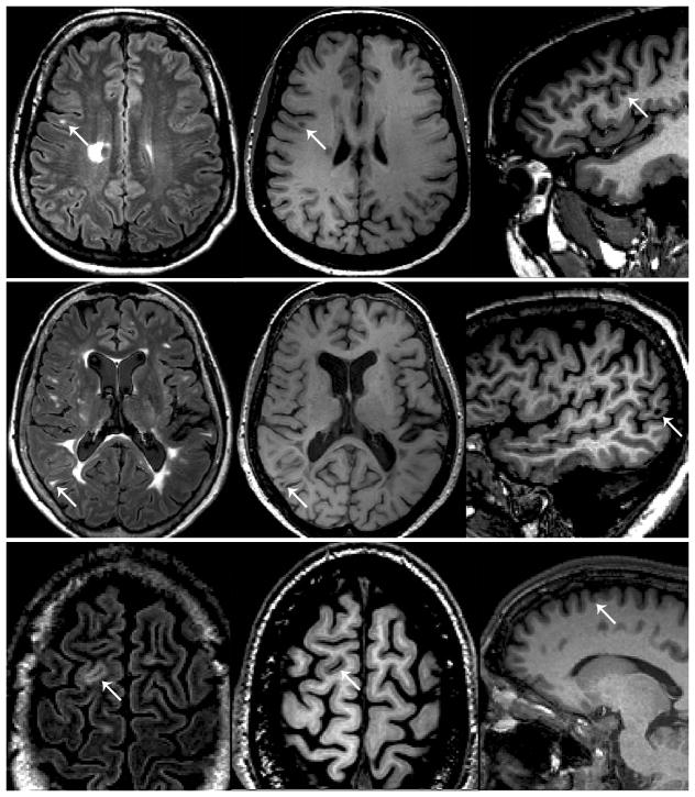
Figure 1.
Examples of lesion classification based on integrated analysis of double inversion recovery (DIR) (left column) and magnetization-prepared rapid acquisition with gradient echo (MPRAGE) (middle and right columns) sequences. Top row: a hyperintense lesion close to the cortex (white arrow) is visible on DIR, but MPRAGE shows that the lesion is located in the white matter. Middle row: a hyperintense lesion close to the cortex (white arrow) is visible on DIR, and MPRAGE shows that the location abuts the cortex (juxtacortical). Bottom row: a hyperintense lesion close to the cortex (white arrow) is visible on DIR, and MPRAGE shows that the lesion is intracortical. Under the proposed system, the lesions in the middle and bottom rows would be classified as “cortical/juxtacortical.”

