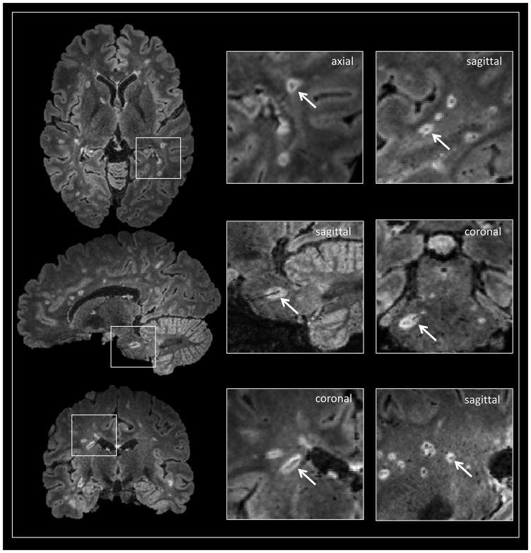
Figure 4.
Noncontrast 3T FLAIR* images (axial, sagittal and coronal views) in a 33-year-old woman with MS. A conspicuous central vessel is clearly visible in the majority of hyperintense lesions. The definition of “perivenular” lesion requires the visualization of the central vessel in at least two perpendicular views (arrows in magnified boxes).

