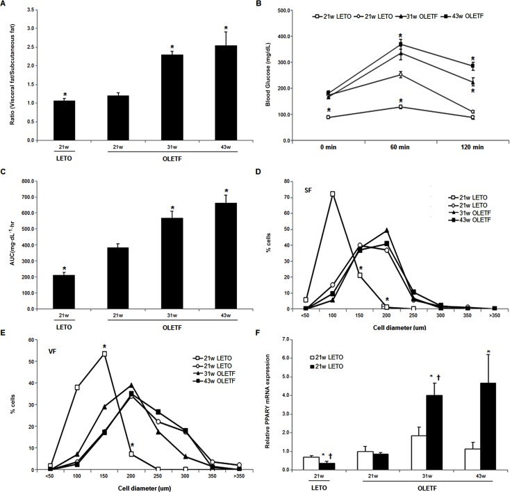
Fig 1.
The effect of aging on the weight of subcutaneous fat/visceral fat ratio (A), OGTT (B), AUC during OGTT (C), adipocyte size distribution of subcutaneous fat (D) and visceral fat (E), and PPARγ2 mRNA levels (F). (*P <0.05 vs. the same deposit in the untreated OLETF rats, †P<0.05 vs. subcutaneous fat in the same group.)

