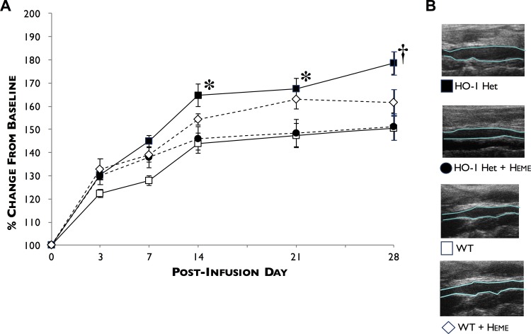
Fig 2. Profiles of AAA development following PPE infusion.
(A) AAA diameters were measured at baseline and at 3, 7, 14, 21, and 28 days post-PPE infusion by ultrasound in WT (white squares), HO-1 Het (black squares), heme-treated WT (white diamonds), and heme-treated HO-1 Het (black circles) mice. (B) Representative longitudinal ultrasound views of AAAs 28-days post-PPE infusion from HO-1 Het, heme-treated HO-1 Het, WT, and heme-treated WT mice. *p<0.015, †p = 0.0025 compared to WT and heme-treated HO-1 Het mice. n = 7 to 11 for each group.

