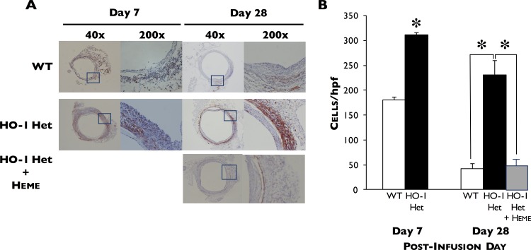
Fig 3. Immunohistochemical staining.
(A) Immunohistochemical staining for Mac1 in aortae harvested from WT and HO-1 Het mice at 7 and 28 days post-PPE infusion and of heme-treated HO-1 Het mice at 28 days post-PPE infusion. (B) Macrophage infiltration in the aortae of WT (n = 4 and 7) and HO-1 Het (n = 4 and 7) mice at 7 and 28 days post-PPE infusion, respectively, and of heme-treated HO-1 Het mice (n = 5) at 28 days post-PPE infusion (right panel). Data shown as cells per high power field (hpf). *p<0.0001 compared to WT mice.

