Fig. 3.
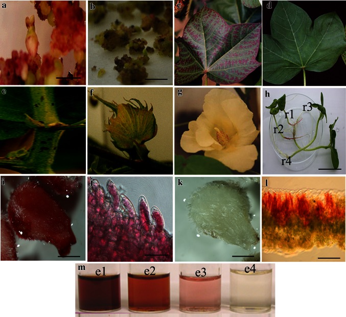
Phenotypic characterization of Lc-transgenic cotton. a Red transgenic calluses and somatic embryos (bar 2.5 mm). b Non-transgenic calluses (bar 2.5 mm). c Transgenic purple colored leaf, indicating anthocyanin accumulation. d Non-transgenic leaf. e Anthocyanin accumulation on stem (red patches). f Transgenic flower bud with a red sepal. g Transgenic flower with a red anther. h T1 siblings showing the segregation of Lc-dependent anthocyanin accumulation in roots. r1 and r2 are positive seedlings with red roots; r3 and r4 are negative seedlings with white roots. i Transgenic immature ovules cultured under light for 1 week (bar 2 mm). Note red fiber cells. j Magnification of J showing a single red fiber cell (bar 10 μm). Note red oily liquid gathered in the central big vacuole. k Transgenic immature ovules cultured in completely dark for 1 week (bar 2 mm). l Transgenic leaf abaxial surface toward the sun; its freehand cross section with red palisade mesophyll cells. The epidermal cells are colorless and transparent (bar 1 mm). m Anthocyanin extract from transgenic dry leaves (e1), transgenic fresh leaves (e2), Wt leaves (e3), and empty control (e4)

