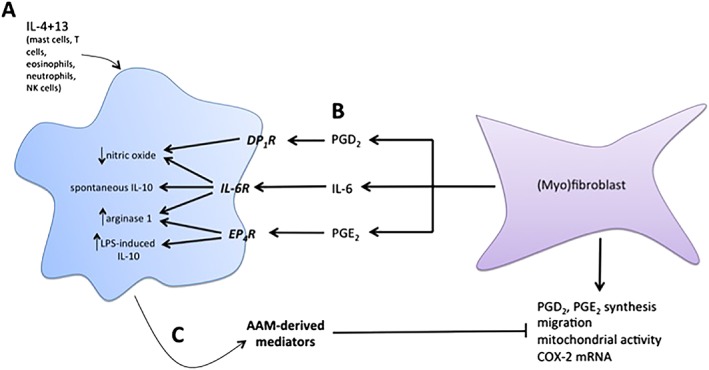
Figure 6.

Model of AAM–myofibroblast interactions. (A) Exposure of macrophages to IL‐4 and IL‐13 induces expression of canonical AAM markers, arginase 1 and Ym1. In the presence of LPS, these AAMs also produce substantial amounts of nitric oxide. (B) Conditioned medium from myofibroblasts, containing IL‐6, PGD2 and PGE2, interacts with specific receptors on macrophages during IL‐4 and IL‐13 priming. In the presence of IL‐6, AAMs display enhanced expression of ARG1 and Ym1, spontaneously produce IL‐10 and suppress LPS‐induced nitric oxide. PGE2 increases ARG1 expression and also enhances LPS‐evoked IL‐10 production, and PGD2 suppresses LPS‐induced NO. (C) Conditioned medium from AAMs induced with MFbCM components suppress PGE2 and PGD2 production from myofibroblasts, and also suppresses migration, mitochondrial activity and COX‐2 gene expression. This indicates that AAMs induced with MFbCM can ‘turn off’ the activity of myofibroblasts, thus demonstrating an important feedback loop that will prevent aberrant activation of both cell types.
