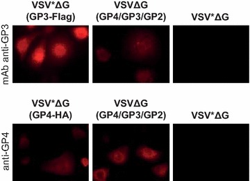
Figure 2.

Characterization of VRP-mediated expression of GP3 and GP4. Immunofluorescence analysis of MARC-145 cells 6 h after infection with VSV*ΔG(GP3-Flag), VSV*ΔG(GP4-HA), VSVΔG(GP4/GP3/GP2) or with the VSV*ΔG control VRP. Expression of GP3 (top panels) and GP4 (bottom panels) were detected with the anti-GP3 mAb VII2D and with a rabbit anti-GP4 serum, respectively.
