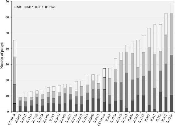
Fig. 3.

Distribution of all counted polyp in three parts of small intestine: SB1-proximal, SB2-middle, SB3-distal and colon. The X-axis represents CC lines, C57BL/6 strain carrying the Apc Min/+ mutation (first black column) and CC-Mean (black column ~ in the middle), while the Y-axis number of polyps. One-way ANOVA performed for statistical analysis, p < 0.05
