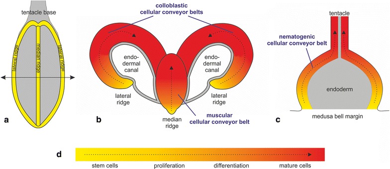
Fig. 1.

Schematic representation of P. pileus and C. hemisphaerica cellular conveyor belts involved in continuous regeneration of tentacle tissues. a Aspect of a P. pileus tentacle root extracted from the animal and viewed from the internal side. The lateral and median ridges (housing stem cells) are highlighted in yellow (rest of tentacle root surface in grey). b Transverse section of the P. pileus tentacle root according to the sectioning plane shown in (a; double arrow) (endoderm in grey). The epithelium of the tentacle sheath that partially surrounds the tentacle root is not represented. c Longitudinal section of a C. hemisphaerica tentacle bulb and its tentacle (endoderm in grey). In b, c the dotted arrowed lines materialise progression along cell lineages and correlative cell movements. d Legend of colour scale used in a–c in terms of dominant cellular stages, according to data published in [46, 49, 50]. Note that cellular conveyor belts are not organised into well-defined compartments (except perhaps for the stem cell niche) but instead form a more or less continuous gradient in terms of the relative abundance of cellular stages
