Fig. 8.

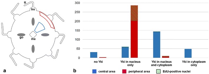
Quantification, in the central versus peripheral area of the C. hemisphaerica exumbrellar epidermis, of observed profiles of anti-Yki staining. a Diagram of the medusa showing approximate localisation of the two areas (blue dotted line central area; red dotted line peripheral area). bu tentacle bulb, go gonad, ma manubrium, tt tentacle. b Graph showing observed numbers of nuclei classified in four categories: no anti-Yki staining; anti-Yki staining in the nucleus but not in associated cytoplasm; anti-Yki staining in nucleus and cytoplasm; anti-Yki staining in cytoplasm but not in nucleus. Blue central area; red: peripheral area. Proportions of nuclei in each category that showed EdU staining are indicated with green hatching. A total of 580 nuclei were counted (282 in the blue zone and 298 in the red zone—raw data given in Additional file 12) using five medusae from the same experiment
