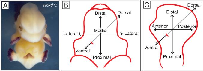
Fig. 1. Comparative organization of the genital tubercle and limb bud.
A. Mouse embryo at E13.5 after in situ hybridization with Hoxd13 antisense RNA probe. Purple stain shows Hoxd13 expression in the handplates, footplates and genital tubercle. B. Major axes of the genital tubercle. C. Major axes of the limb.

