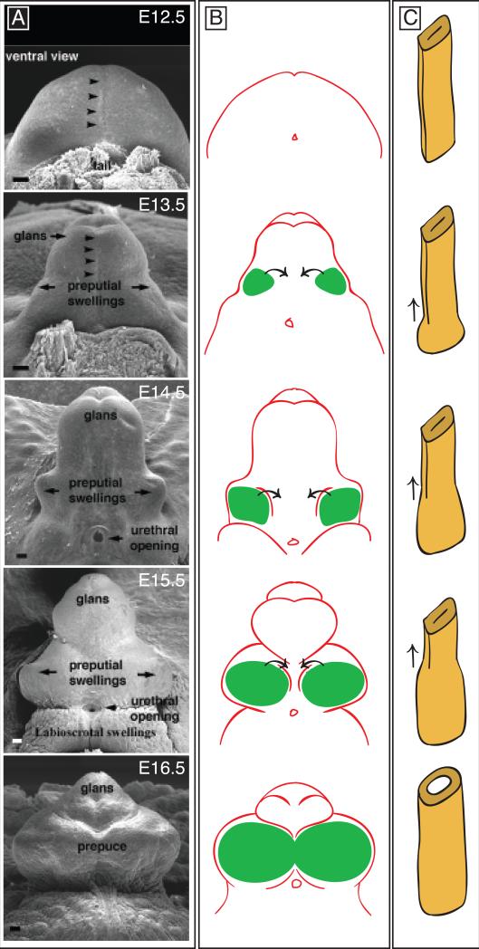
Fig. 2. Development of the external genitalia.
A. Scanning electron micrographs of mouse genital tubercles from E12.5-E16.5. All panels are ventral views. Arrowheads mark the position of the urethral plate along the ventral midline. Modified from Perriton et al., 2002. B. Development of the prepuce. Paired preputial swellings are shown in green. Ventral and lateral growth of the preputial swellings results in development of a prepuce that surrounds the glans of the penis or clitoris. C. Schematic diagram of urethral tube formation. At E12.5, the endodermally-derived urethral epithelium is a bilaminar plate without a lumen. Tubulogenesis then progresses from proximal to distal (arrows), resulting in conversion of the closed plate to an open tube.

