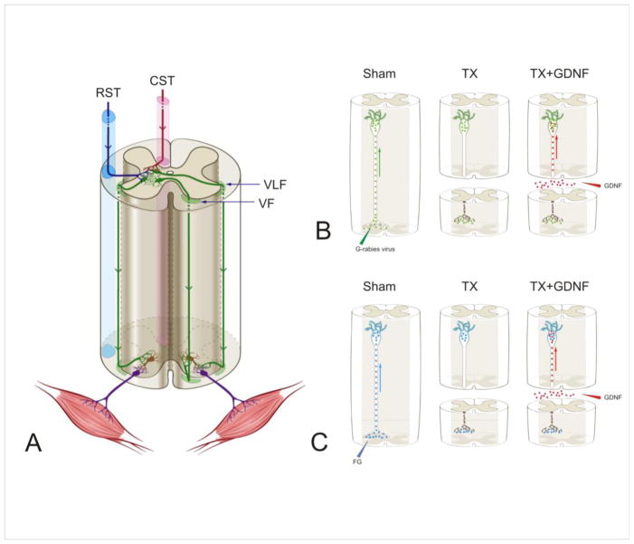
Figure 1.
(A) Schematic diagram of the descending propriospinal tract system (dPST). dPST pathways descending several spinal segments are located in both the ventral and ventral lateral funiculi (VF and VLF, respectively). The dPST projects through either contralateral or ipsilateral VF or VLF and innervates motoneuron pools directly or indirectly through interneurons. dPST neurons receive convergent supraspinal innervation, including those from the corticospinal (CST) and rubrospinal (RST) tracts. Descending propriospinal neurons are indicated in green, interneurons in brown and motoneurons in purple. (B, C) Schematic diagrams of the experimental designs for the dendritic morphology study (B) and the neurotransmitter study (C). The three figures in each panel, from left to right, show propriospinal neurons were first retrogradely infected by a G-mutated rabies virus (green particles) that expressed green fluorescence protein which filled the dendritic compartments of dPST neurons (B) or retrogradely labeled by FluoroGold (FG) (blue particles) (C). Spinal cords then received either a transection injury or a transection + glial cell line-derived neurotrophic factor (GDNF) (red dots) applied to the lesion site to be retrogradely transported to the soma.

