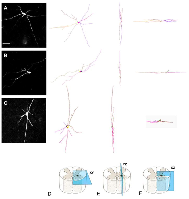
Figure 2.
Camera lucida reconstructions of three propriospinal neurons (rows A, B, C) with different dendritic patterns as if viewed in the transverse (D), parasagittal (E), and horizontal (F) planes. Row A shows a dPSN that extended its dendritic branches in medial, lateral, ventral and dorsal directions. Row B shows a dPSN that extended its dendritic branches predominantly in medial and lateral directions. Row C shows a dPSN that has more dendritic branches extending in ventral and dorsal directions. Scale bar: 100μm.

