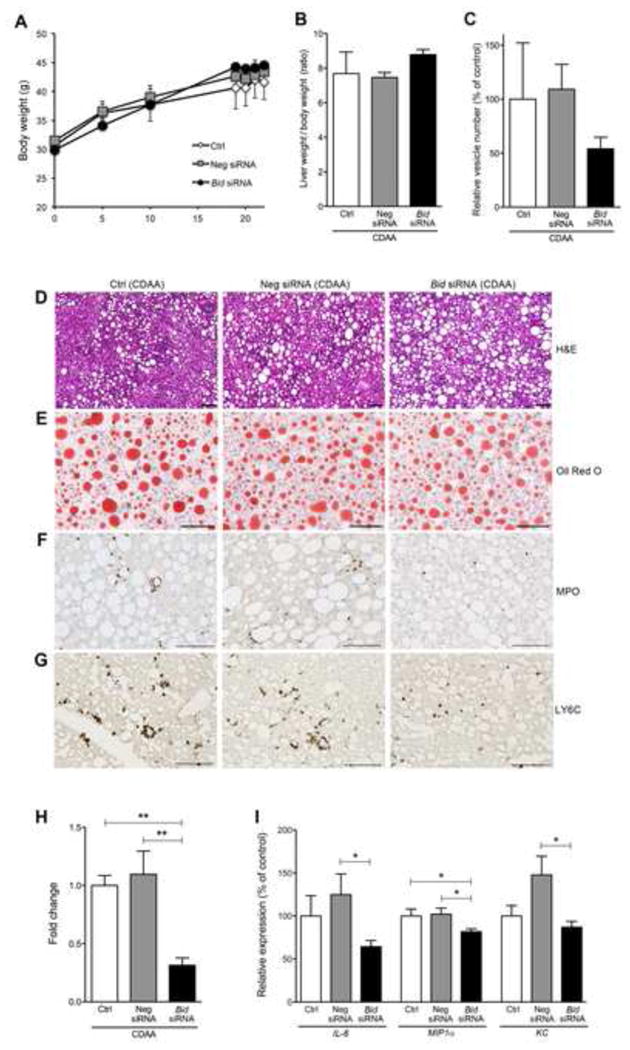
Fig. 2. Bid knockdown reduces circulating levels of extracellular vesicles and improves inflammation independent of steatosis in mice fed a CDAA diet.

The effect of Bid suppression in (A) body weight (B) ratio of liver weight / body weight, and (C) circulating extracellular vesicles in mice fed a CDAA diet administered with control, Neg siRNA, or Bid siRNA. Liver histology of Bid suppression revealed no change in steatosis, but did show reduced liver inflammation in comparison to control (D-G). (D) Haematoxylin-eosin staining of liver sections in mice fed a CDAA diet administered with control, Neg siRNA, or Bid siRNA. Scale bar, 100 μm. (E) Oil Red O staining of liver sections in mice fed a CDAA diet administered with control, Neg siRNA, or Bid siRNA. Scale bar, 100 μm. (F) Immunohistochemical staining specific for MPO (neutrophils) (F) or Ly6C (G) of liver sections in mice fed a CDAA diet administered with control, Neg siRNA, or Bid siRNA. Scale bar, 100 μm. (H) Bar graph shows quantification of MPO positive cells. ** p<0.01. (I) Gene expression of inflammatory genes as measured by qPCR. All gene expression levels were normalized to housekeeping control, β2 microglobulin, and shown relative to the expression levels of mice fed a CDAA diet administered with control. *p<0.05. Values are mean ± SEM. Ctrl: Control, Neg siRNA: negative siRNA
