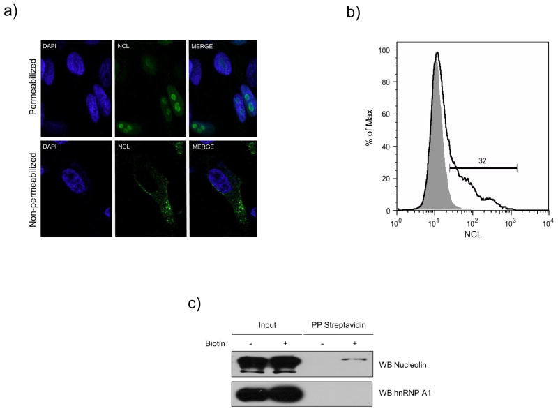
Figure 1.
Nucleolin is expressed in the surface of CrFK cells. A) Non-permeabilized and permeabilized CrFK cells with acetone, were stained with an anti-NCL antibody (green), and visualized by indirect immunofluorescence using Alexa 488 as secondary antibody. DAPI was used for nuclei staining (blue). B) Non-permeabilized CrFK cells were incubated with anti-NCL (black line) or anti-rabbit IgG antibodies (gray shaded area), used as an isotype control. C) CrFK cells were treated with sulfosuccinimidyl-6-(biotin-amide) hexanoate (+) or vehicle (−) and biotinylated proteins were precipitated with streptavidin-agarose, and separated by SDS-PAGE. NCL and hnRNP A1 proteins were detected by immunoblotting using specific anti-NCL and anti-hnRNP A1 antibodies respectively.

