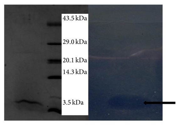
Figure 1.

Purified fused pediocin analyzed by SDS-PAGE. Gel was removed and cut into two parts. One half, containing molecular weight marker (lane 2) and the purified pediocin, was stained with CBB R-250. The other half, containing the purified bacteriocin (lane 3), was overlaid with Staphylococcus aureus and incubated at 37°C for 16 h. Arrow indicates bacteriocin activity.
