Fig. 3.

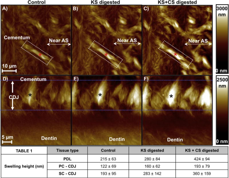
AFM micrographs illustrated a change in swelling characteristics of a single PDL-insert within cementum (A–C, marked by a red asterisk) and in the CDJ (D–F, marked by a black asterisk) following GAG digestion. The intensity scale bars on the right for each row illustrate variation in topographical height. (A, D) exhibited swelling equivalent to undigested conditions, (B, E) post KS-digestion swelling, and (C, F) post KS + CS digestion swelling. The averaged results (Table 1) of PDL-inserts revealed a significant increase in swelling upon GAG digestion (ANOVA tests (P < 0.05)). It should be noted that the same PDL–cementum and cementum–dentin interfaces sites were analyzed for each KS and CS digestion condition to accurately monitor the effect of digestion on swelling and mechanical properties. AS: Attachment site.
