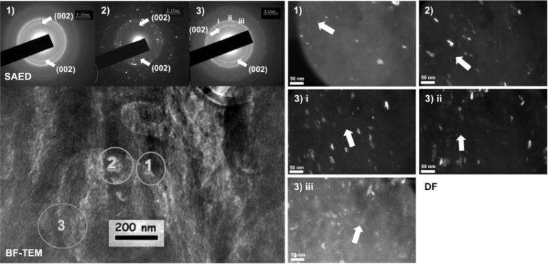
Fig. 5.

Left: bright-field (BF) TEM image with insets of selected area electron diffraction (SAED) patterns at regions marked 1–3. Right: corresponding dark-field (DF) images highlighted the orientation of hydroxyapatite crystals along the 002 c-axis, which coincided with collagen fibril direction indicating the presence of intrafibrillar mineral. While crystal orientation maintained a close match to fibril direction, SAED and DF indicated a spread in crystal orientation within the sampled regions and a strong possibility of the presence of extra-fibrillar mineral.
