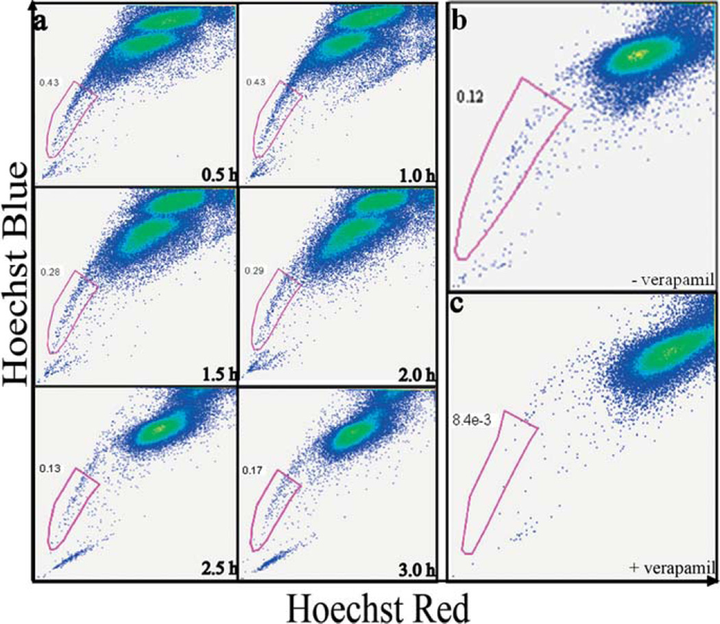
Figure 2.
CICs can be isolated from the cell line MX-1 using Side Population method. (a) Fluorescence activated cell sorting (FACS) dot plots showing CICs isolation from MX-1 cancer cell line at various time points. MX-1 cells were incubated with Hoechst 33342 nuclear dye for 0.5–3 hrs. The x-axis ‘Hoechst Red’ and y-axis ‘Hoechst Blue’ is the fluorescence at 675 nm and 450 nm respectively. At 2.5 hrs, the majority G0-G1-S population was saturated with the dye and the separation of the low and high fluorescence cells is evident. The low fluorescent population is collected as CICs. Verapamil, a molecular pumps inhibitor added along with Hoechst 33342 during incubation. FACS dot plots of MX-1 analysed without (b) and with (c) verapamil. Verapamil abolishes the side population, demonstrating that CICs can efflux dyes only through molecular pumps.

