
Fig. 1.
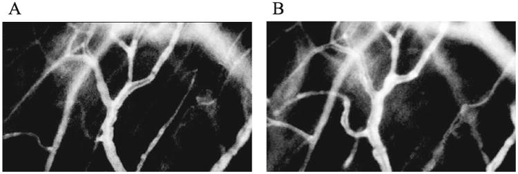
Fluorescent image of C3H mouse skin as recorded during intravital microscopic examination before irradiation (panel A) and at one day after 50 Gy irradiation (panel B). The same vessels were observed for each experiment before and after irradiation. The total volume and total surface area of the vessels were measured using the fluorescent image.