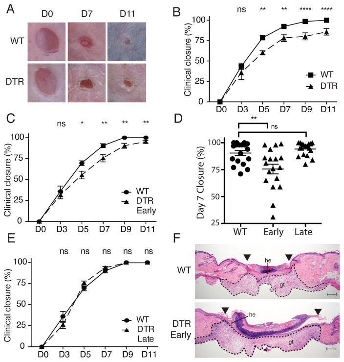
Figure 1. Tregs facilitate skin wound repair.
Foxp3-DTR or WT mice were treated with DT 2 days prior to full thickness wounding of dorsal skin and every 2 days thereafter. (A) Representative images of wounds at specific times after injury (D, day). (B) Mean percentage of wound closure with time after injury. (C) Mean percentage of wound closure with time after injury between WT and Foxp3-DTR mice treated with DT “early” after wounding. (D) Representative plot of mean percentage of wound closure 7 days after wounding. Each symbol represents an individual wound. (E) Percent of wound closure with time after injury between WT and Foxp3-DTR mice treated with DT ‘late’ after wounding. (F) Representative histology of skin wounds at day 7 post-injury between WT and Foxp3-DTR mice treated ‘early’ with DT. Arrowheads denote wound edges; he, hypertrophic epithelium; gt, granulation tissue; scale bars, 200μm. Representative data is shown from ≥ 3 replicate experiments with ≥ 3 mice per group. Error bars in all panels represent the mean ± SEM. ns p>0.05, * p≤0.05, ** p≤0.01, *** p≤0.001, **** p≤0.0001.

