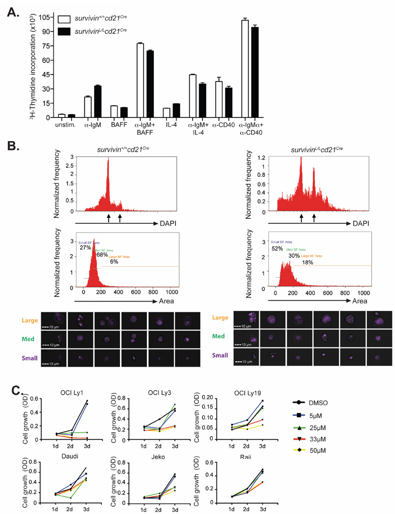
Figure 6. Survivin-deficient B cells accumulate aberrant levels of DNA after mitogenic stimulation.
(A) B cells from survivinL/Lcd21Cre and survivin+/+cd21Cre mice were cultured unstimulated or in the presence of the indicated stimuli for 3 days. Graphs show 3H-thymidine incorporation. The following concentrations were used: 100ng/mL IL4; 25ng/mL BAFF, 5µg/mL anti-CD40, 10µg/mL anti-IgM (B) Morphology and DNA content of LPS (10 µg/mL) plus IL-4 (10 ng/mL) stimulated survivinL/Lcd21Cre and survivin+/+cd21Cre B cells on day 3 of culture was analyzed using an ImageStream Imaging Flow Cytometer. Arrows indicate 2n and 4n DNA content. Results are representative for 2 independent experiments. (C) Cell growth of OCI-Ly1 (GCB-DLBCL), OCI-Ly3 (ABC-DLBCL), OCI-Ly19 (GCB-DLBCL), Daudi (Burkitt‘s lymphoma), JeKo (Mantle cell lymphoma), Raji (Burkitt‘s lymphoma) cells 1, 2 and 3 days after the beginning of cell culture in the presence of the indicated concentration of the survivin inhibitor S12 was determined using a colorimetric cell counting assay. Displayed are the obtained OD values minus the value of a blank sample. The results are representative of three independent experiments.

