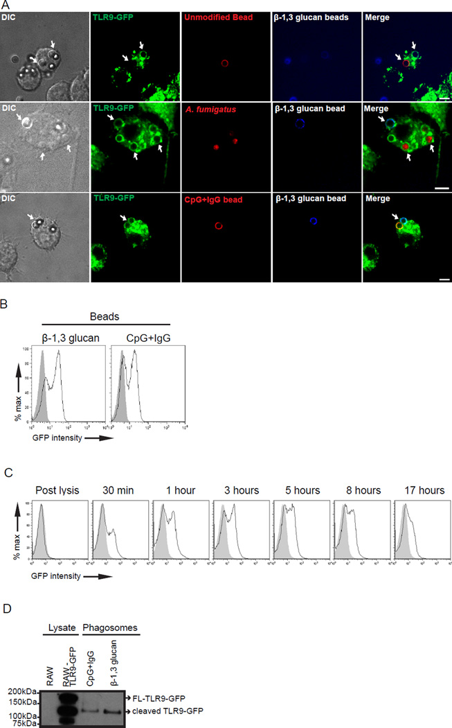
Figure 1.
TLR9 is specifically recruited to β-1,3 glucan phagosomes. (A) Confocal microscopy of RAW macrophages expressing TLR9-GFP (green) incubated with Alexa fluor 647-labeled β-1,3 glucan beads (blue) and Alexa fluor 568-labeled polystyrene beads (red-top panel), or A. fumigatus-RFP (red-middle panel), or Alexa fluor 568-labeled CpG-IgG beads (red-bottom panel) for 30 min. Size bar indicates 5µm. Original magnification X60 or X100. (B) Purified phagosomes containing either β-1,3 glucan beads or CpG-IgG beads were assessed for TLR9-GFP recruitment (black line) by phagoFACS and compared with polystyrene beads control (gray shaded histogram). (C) Kinetics of TLR9-GFP recruitment (black line) to purified β-1,3 glucan-containing phagosomes were assessed by phagoFACS for the indicated times and compared to polystyrene bead-only control (gray shaded histogram). β-1,3 glucan beads were added to cell lysates assessed for TLR9-GFP recruitment (D) β-1,3 glucan beads and CpG-IgG beads were incubated with 2x10⁶ RAW TLR9-GFP cells for 3 h. Lysate control and 1x10⁶ purified phagosomes were analyzed by immunoblot and blotted for GFP. Arrows indicate full-length-TLR9GFP (FL-TLR9-GFP) and cleaved TLR9-GFP as labelled. Data are representative of five independent experiments.

