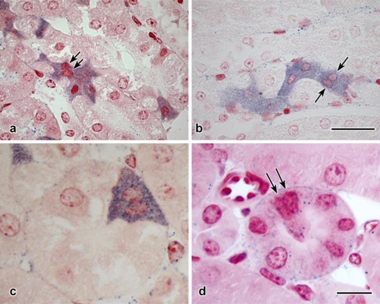
Fig. 3. Configurations of S3 segment proximal tubular cells.
a, b: Two-micrometer paraffin sections, NBT-perfused and Neutral Red counterstained. In both micrographs, diformazan-stained cells are found in groups, and in some cases (arrows) doublets of nuclei are closely apposed.
c: Detail of cross-sectioned pars recta tubule. The diformazan-positive profile contains two nuclear profiles.
d: Semithin (<0.25 μm) plastic section, glutaraldehyde-fixed, unosmicated tissue with basic fuchsin counterstaining. A characteristic cell is identifiable by the deposition of diformazan particles (cf. Fig. 2 i-k), and a doublet of nuclei, similar to that seen in c, is visible within its confines (arrows).
Scale bar in b = 25 μm and applies to a and b; scale bar in d = 10 μm and applies to c and d.

