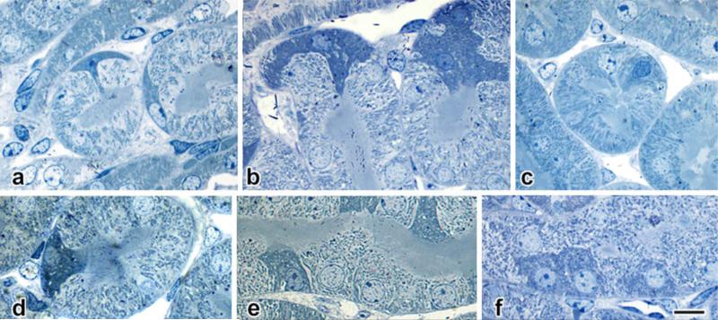
Fig. 5. Structural details in semithin (<0.25 μm) plastic sections of glutaraldehydeperfused kidney.
In the same animal (42-day sham-operated male mouse), a spectrum of morphologies is found, ranging from seemingly surface-applied cells with cytoplasmic processes projecting inward between normal epithelial cells (a, b) to cells with dark nuclei (c, d) and profiles that occupy the full depth of the tubule wall, to cells with larger nuclei (e, f) that more closely resembling those of their neighbors, but retaining their cytoplasmic osmiophilia. Scale bar in f = 10 μm and applies to all panels.

