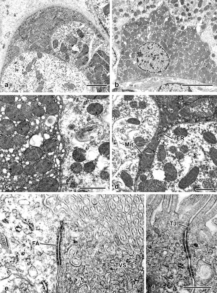
Fig. 6. Fine-structural details of glutaraldehyde-perfused mouse kidney.
a. An electron-opaque profile forms part of this tubule's surface (compare with the semithin section image in Fig. 5a). Its cytoplasm is filled with mitochondria.
b. Survey view, showing an ovoid, heterochromatic nucleus and cytoplasmic concentration of mitochondria.
c. Apposition of two S3 epithelial cells, demonstrating higher mitochondrial concentration and ribosome-rich cytoplasm of one (at left) as opposed to an adjacent S3 segment epithelial cell containing scattered mitochondria and electron-lucent cytoplasm typical of the majority of proximal-tubule epithelial cells.
d. Thin cytoplasmic process extending between two proximal tubular epithelial cells. Both mitochondria (Mit) and rough endoplasmic reticulum (rER) are present within the confines of the process.
e. Apical portions of an S3 segment epithelial cell (at left) and a mitochondrion-rich cell (at right), with a prominent “intermediate” junction (fascia adherens: FA) joining the two cells. Both the brush border and tubulovesicular system elements (TVS) are evident.
f. Apical junctional complex formed between two adjoining mitochondrion-rich cells, with both a tight junction (TJ) and adherens junctions.
Scale bar in a = 2 μm; in b = 5 μm; in c = 1 μm; in d = 1 μm; in f = 0.5 μm and applies to e and f.

