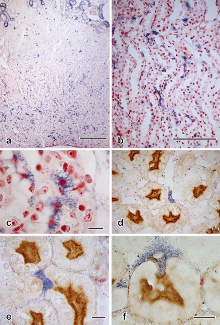
Fig. 7. Extratubular cells positive for NBT reduction.
a. Medulla in 10-μm section (cortex shown in Fig. 2a). Multiple diformazan-positive cells are present.
b. Two-micrometer section from medullary region shown in A, showing distribution of cells in the medullary tubules.
c. Detail of b, illustrating the dendritic profiles and densely staining nuclei of diformazan-stained tubule cells.
d. Two-micrometer section (Lotus staining) with a blue cell constituting part of the wall of a portion of the thin loop.
e. Interstitial diformazan-positive cell sandwiched among several pars recta profiles.
f. A diformazan-positive cell appears to adhere to the outer border of a proximal tubule while inserting a cytoplasmic projection into the tubule. Lotus staining with Neutral Red counterstain. Scale bar in a = 250 μm; scale bar in b = 100 μm; scale bars in c-f each =10 μm.

