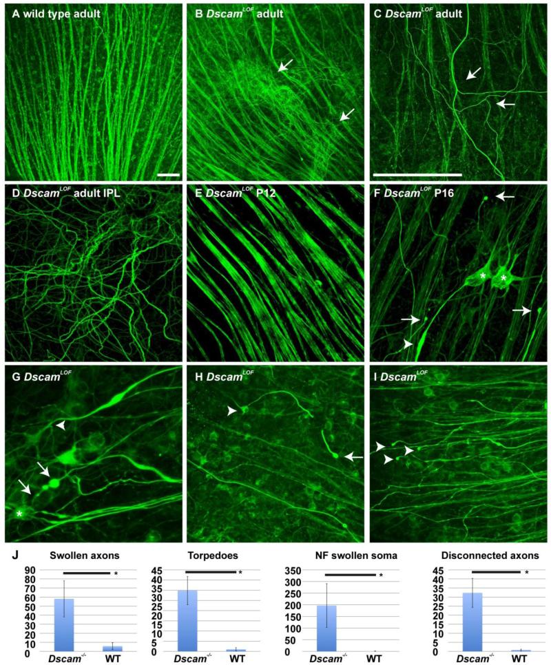
Figure 1. Postnatal axon branching and stress in DscamLOF mice.
Wild type and DscamLOF retinas stained with antibodies to neurofilament medium chain protein. A and B, Abnormal projection of retinal ganglion cell (RGC) axons was observed in the DscamLOF retina (B: arrows). C, Abnormal axon branching was observed in the peripheral retina (arrows). D, Abnormal axons were observed to project through the inner plexiform layer (IPL) of the DscamLOF retina. E, Axon morphology in DscamLOF mice was normal prior to eye opening, which occurs after postnatal day 12 (P12). F, Signs of axon stress, including accumulation of neurofilament protein in the cell soma (asterisks), blebbing of axons (arrows), and swollen axon torpedoes (arrow head), were observed in DscamLOF mice by P16. G, Swollen axons with torpedoes (arrows) and branching (arrowhead). H, RGC (arrowhead) with a broken axon that terminates in a bleb (arrow). I, Axons not visibly connected to RGC soma that terminate in blebs. N>3 at all ages. J, The number of swollen axons, axon torpedoes, neurofilament (NF) swollen somata and disconnected axons were significantly increased in DscamLOF retinas compared to wild type controls at P16 (Student's t-test < 0.01). A-F Dscamdel17, G-I Dscam2J and sibling controls used. N≥6. Scale bar (in A) = 50 μm; A, B, (in C) = 25 μm C-F, (in C) = 50 μm; G, (in C) = 100 μm; H and I.

