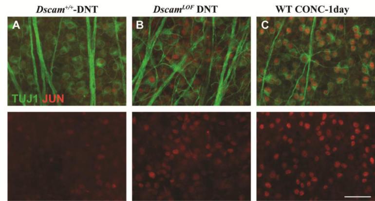
Figure 7. JUN upregulation in DscamLOF mice.
Retinas were stained with antibodies to JUN and TUJ1. A, A few JUN positive cells are observed in wild type retinas. B, A large number of RGCs are JUN-positive in the DscamLOF retinas. C, Wild type retina showing the upregulation of JUN in all RGCs after an acute axonal insult, optic nerve crush. N>3. The scale bar is equivalent to 50 μm.

