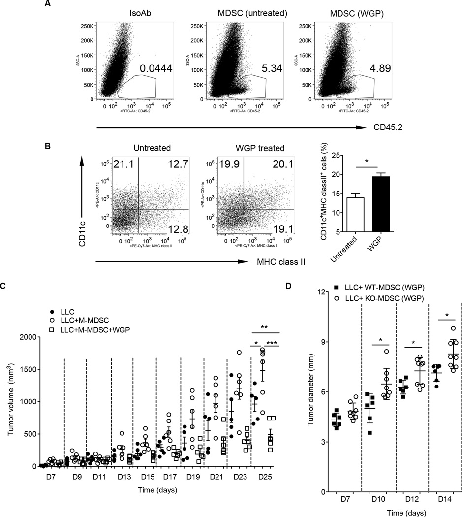
Figure 7. Particulate β-glucan treatment induces M-MDSC differentiation in vivo with reduced tumor growth.
Tumor M-MDSC sorted from C57Bl/6 LLC tumor-bearing mice (CD45.2) were treated with or without WGP for overnight and then intratumorally injected into SJL tumor-bearing mice (CD45.1). Mice were sacrificed after 7 days and single cell suspensions from tumors were stained with anti-CD45.2 or isotype control mAb (A) and CD11c and MHC class II. Cells were gated on CD45.2+ cells (B). The percentage of CD11c+MHC class II+ cells was summarized. (C) M-MDSC sorted from LLC tumor-bearing mice were treated with or without WGP and then mixed with LLC cells for injection. LLC alone was used as control. Tumor growth was monitored and recorded. (D) M-MDSC sorted from LLC tumor-bearing wildtype or dectin-1 KO mice were treated with WGP and mixed with LLC cells for injection. Tumor growth was monitored. *p<0.05, **p<0.01, ***p<0.001.

