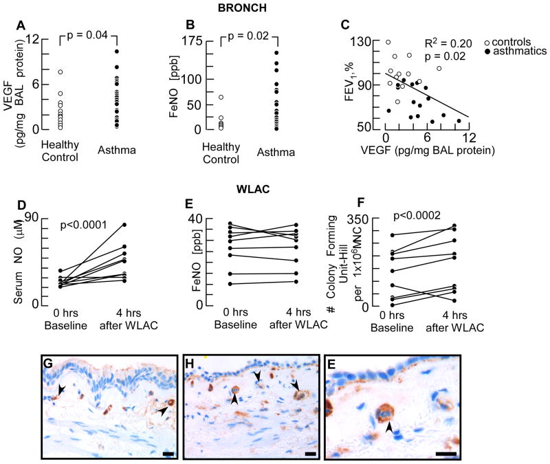
Fig 1. Early Endothelial Activation after Allergen Exposure in Human Asthma.
[A] VEGF levels in BAL fluid measured by ELISA. [B] Fraction of exhaled NO (FeNO) in asthma and healthy control subjects. Each dot represents data from one subject. [C] Correlation between %FEV1 and BAL VEGF levels. Open dots are values from healthy controls and filled dots from asthma patients. [D – F] Serum and exhaled NO levels, and circulating proangiogenic hematopoietic progenitors before and after whole lung allergen challenge (WLAC). [G-E] Immunohistochemistry of CCR3 expression in submucosal endothelium. Healthy control [G] and asthmatic [H-E] human bronchi showed CCR3 expression on endothelial cells in the bronchial capillaries. High power image [E] showed CCR3 positive capillary endothelium with an inflammatory cell in the lumen. Scale bar = 200 μm

