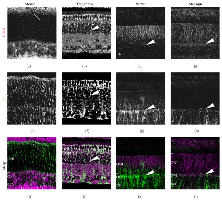
Figure 4.
CB2R immunoreactivity in Müller cells. Vertical sections from the mouse retina (first column), tree shrew retina (second column), vervet retina (third column), and macaque retina (fourth column). Confocal micrographs of coimmunolabeling for CB2R and the cell-type-specific marker for glial Müller cells, glutamine synthetase (GS). Each protein immunofluorescent signal is presented alone in grayscale: CB2R in the first line and GS in the second line; then the two are presented merged (third line: CB2R in magenta and GS in green). Arrowheads point to Müller cell processes that all express CB2R, except in mice (arrows). ONL: outer nuclear layer; OPL: outer plexiform layer; INL: inner nuclear layer; IPL: inner plexiform layer. Scale bar = 30 μm.

