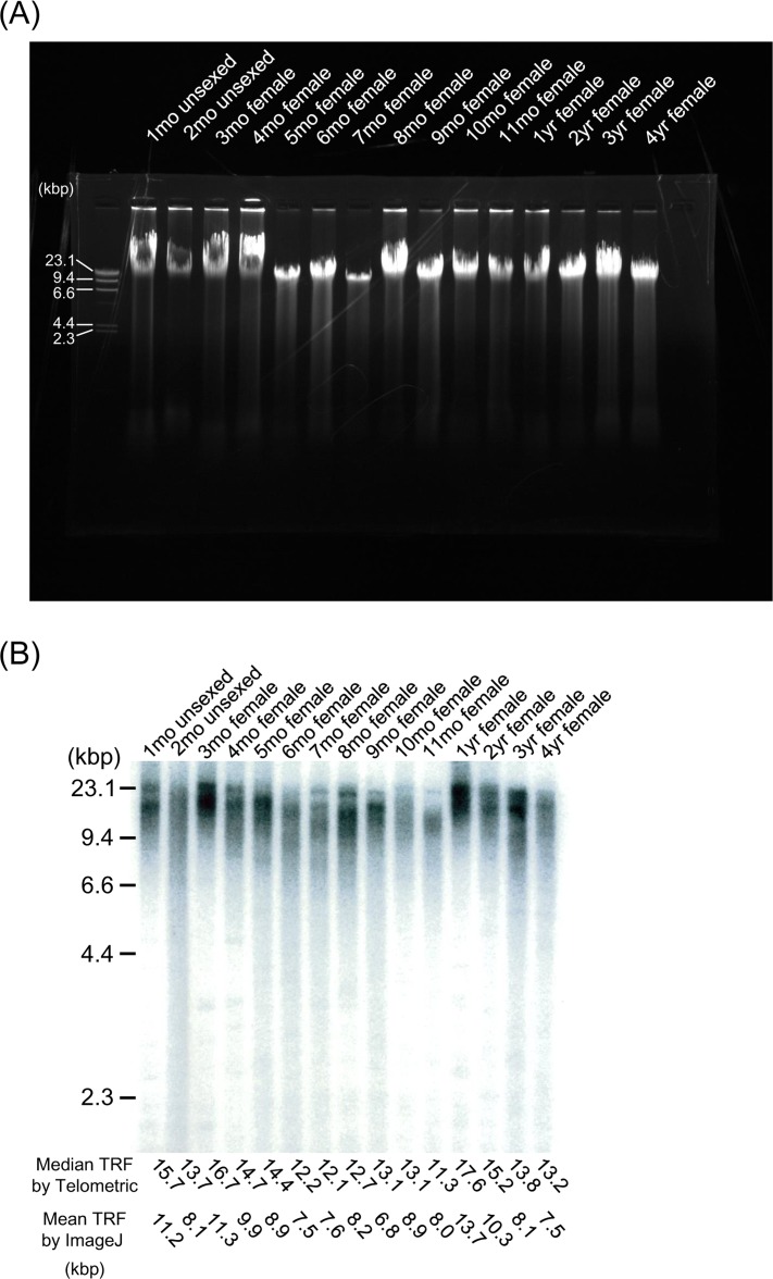
Figure 2. TRF length from embryo to extreme old age in the medaka.
(A) Representative gel electrophoresis analysis using genomic DNA from the whole body at different ages. DNA samples were resolved on a 0.8% (wt/vol) agarose gel at 100V for 30 min. All lanes contain DNA larger than the 23.1-kbp marker, and appear as single compact crowns that have migrated in parallel as an undegraded intact sample. (B) Representative Southern blot analysis using genomic DNA from the whole body at different ages. The TRF of telomeric DNA yielded wide smears in all lanes. TRF values (median values determined using Telometric version 1.2 and mean values determined using ImageJ version 1.39) (kbp) are listed at the bottom. (C) Scatterplots of telomere dynamics obtained using Telometric and the fitted regression model. G (dark gray shading), growth stage; Ado (clear), adolescent stage; Adu (light gray shading), adult stage.


