
FIG. 13.
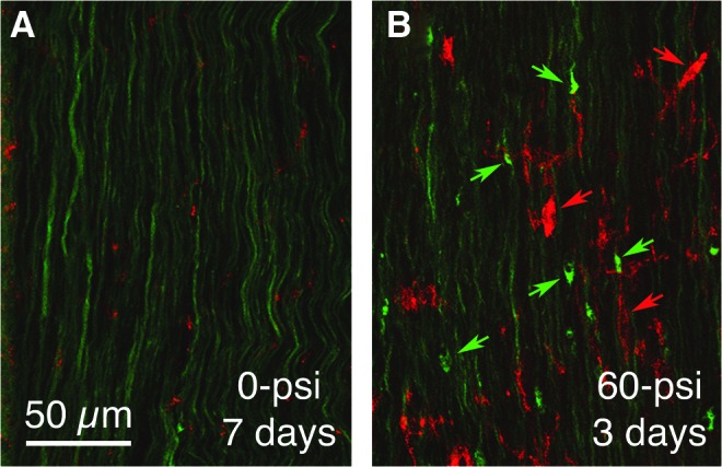
Immunostaining for the activated microglial marker OX6 (red) in right optic tract of a 60-psi blasted enhanced yellow fluorescent protein (EYFP)-reporter mouse (B) compared with a sham blasted EYFP-reporter mouse (A), 3 days after blast. The EYFP labeling is green in both. Note the axon bulbs in B (green arrows) and OX6+ microglia (red arrows). Color image is available online at www.liebertpub.com/neu