
FIG. 9.
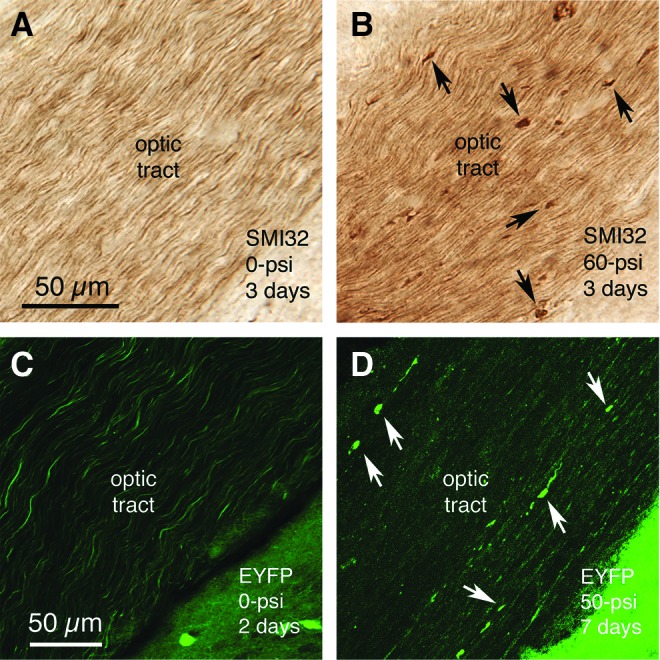
Swollen axon bulbs form in the optic tract by 2–7 days after 50–60 psi blasts, as visualized by SMI-32 immunostaining of optic nerve axons (A, B), or enhanced yellow fluorescent protein (EYFP) labeling in reporter mice (C, D). Note that more bulbs are evident in the SMI-32-immunostained material, because many fewer optic tract axons are EYFP-labeled in the reporter mice than are immunostained for SMI-32. Some of the axon bulbs in B and D are indicated by arrows. Images A and B are at the same magnification as one another, and images C and D are at the same magnification. Color image is available online at www.liebertpub.com/neu