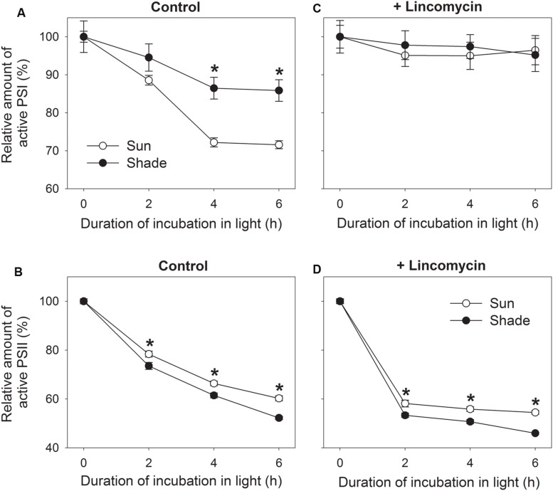
FIGURE 2.
Relationship between photoinhibition of PSII (B,D) and PSI (A,C) in sun and shade tobacco leaves. Detached leaves incubated in the presence or absence of lincomycin (1 mM) overnight in darkness were exposed to 4°C and 300 μmol photons m-2 s-1 for 2, 4, or 6 h. Fm was measured after dark adaptation to estimate the amount of active PSII centers. Pm was measured after dark adaptation to estimate the amount of active PSI centers. All values are expressed relative to the controls before chilling-light treatment, and shown as means ± SE (n = 6). Asterisks indicate significant differences between the sun and shade leaves.

