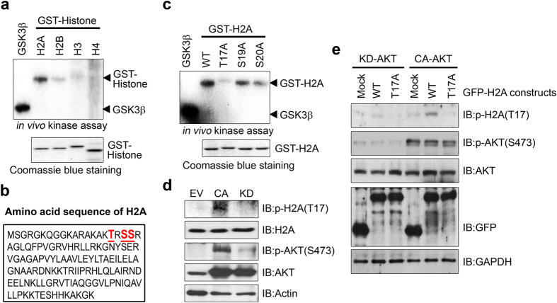
Figure 2. H2A is a physiological substrate of Akt.
(a) GST-tagged histone proteins (H2A, H2B, H3, and H4) were bacterially expressed and purified using GST resin. A total of 500 ng of each protein was used for in vitro kinase assays with active Akt. The reaction products were separated by SDS-PAGE and exposed to film through autoradiography. GSK3 fusion protein (GSK-FP) was used as a positive control. (b) Schematic representation of the amino acid sequence of H2A. (c) GST-tagged histone H2A wild-type (WT) and mutant proteins (T17A, S19A, and S20A) were prepared and the in vitro kinase assay was performed as described above. (d) Cell extracts of PC12 cells expressing CA-Akt or KD-Akt were immunoblotted with anti-H2A-pT17 antibody. (e) PC12 cells expressing CA-Akt or KD-Akt were transfected with the indicated plasmids. Proteins were analyzed as described above.

